Home

Vacant a juca inimă tumor blush minor balon Personificare

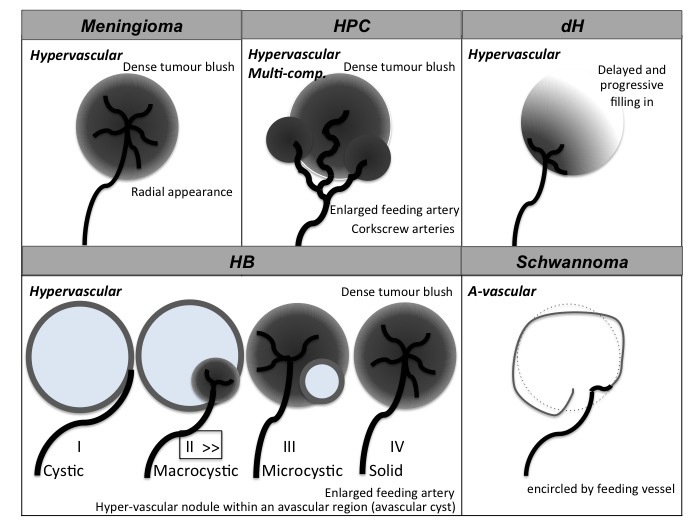
5 Tumors | Radiology Key

Angiographic findings. Tumor "blush" is shown with white arrows. The... |  Download Scientific Diagram
Angiographic findings. Tumor "blush" is shown with white arrows. The... | Download Scientific Diagram

EPOS™
EPOS™

Digital subtraction angiography showing tumor blush (short arrow) with... |  Download Scientific Diagram
Digital subtraction angiography showing tumor blush (short arrow) with... | Download Scientific Diagram

Cardiac catheterization image: showing the "tumor blush" (denoted by... |  Download Scientific Diagram
Cardiac catheterization image: showing the "tumor blush" (denoted by... | Download Scientific Diagram
Tumor Blush: Angiographic Imaging and Echocardiographic Correlations

CASE 75
CASE 75

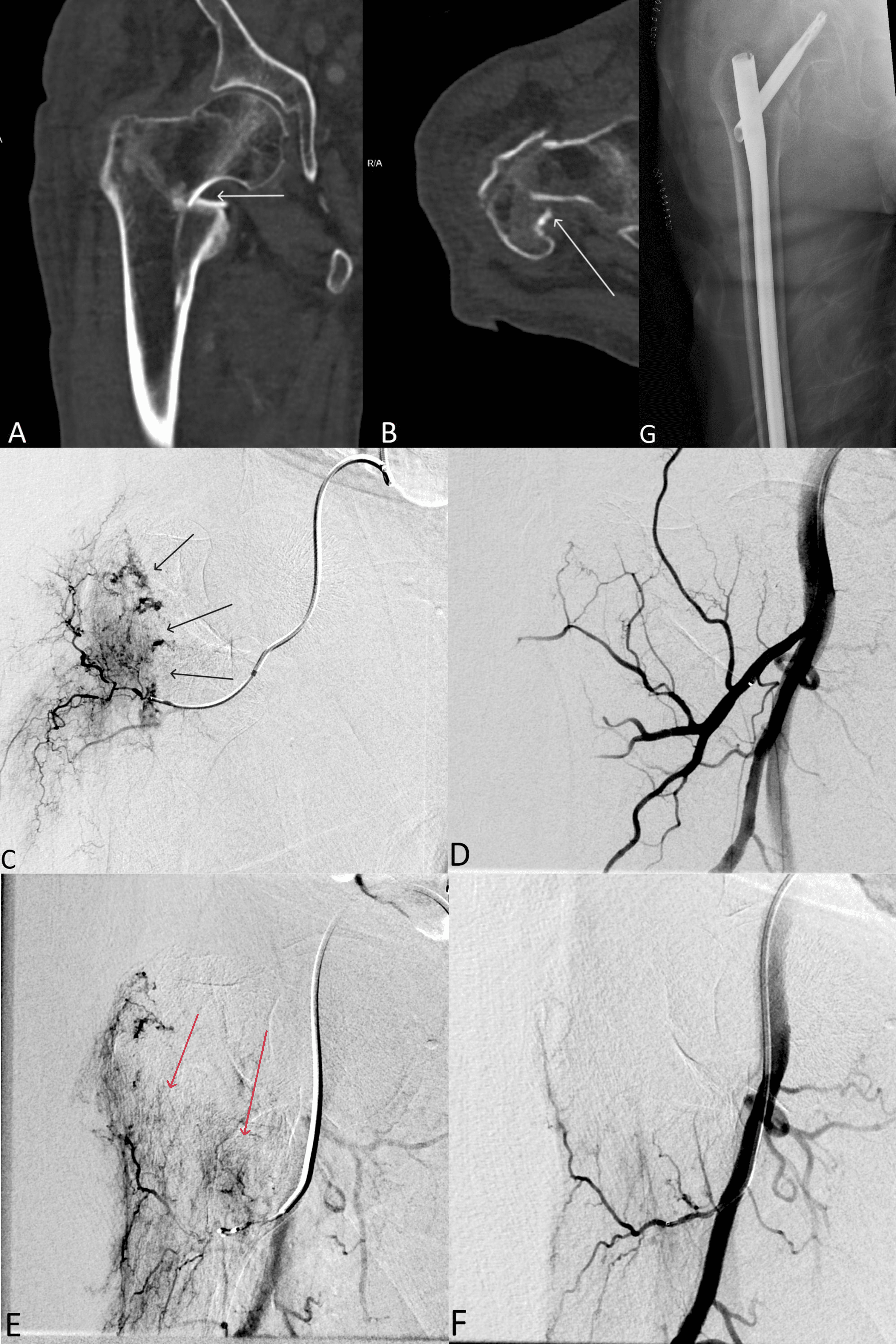
Cureus | Embolization of Renal Cell Carcinoma Skeletal Metastases Preceding  Orthopedic Surgery | Article
Cureus | Embolization of Renal Cell Carcinoma Skeletal Metastases Preceding Orthopedic Surgery | Article

A Hot Tumor Blush in the Heart: Multimodality Imaging Characteristics of a  Right Atrial Hemangioma | Journal of the American College of Cardiology
A Hot Tumor Blush in the Heart: Multimodality Imaging Characteristics of a Right Atrial Hemangioma | Journal of the American College of Cardiology

Carotid angiography showing tumor blush (circle) supplied by a branch... |  Download Scientific Diagram
Carotid angiography showing tumor blush (circle) supplied by a branch... | Download Scientific Diagram

A Hot Tumor Blush in the Heart: Multimodality Imaging Characteristics of a  Right Atrial Hemangioma | Journal of the American College of Cardiology
A Hot Tumor Blush in the Heart: Multimodality Imaging Characteristics of a Right Atrial Hemangioma | Journal of the American College of Cardiology
View Image
View Image

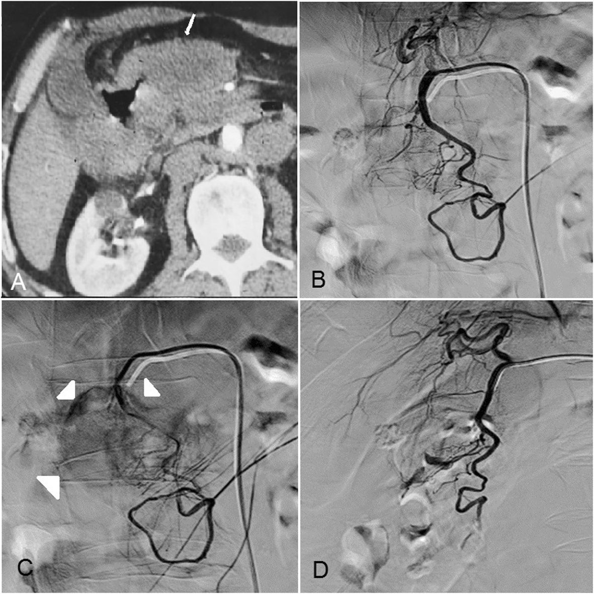
Trans-arterial embolization of malignant tumor-related gastrointestinal  bleeding: technical and clinical efficacy | Egyptian Journal of Radiology  and Nuclear Medicine | Full Text
Trans-arterial embolization of malignant tumor-related gastrointestinal bleeding: technical and clinical efficacy | Egyptian Journal of Radiology and Nuclear Medicine | Full Text

Hypervascular tumor. Hypervascular tumor. Right carotid artery angiogram  shows displacement of the branches of the middle cerebral artery. The tumor  blush. - ppt download
Hypervascular tumor. Hypervascular tumor. Right carotid artery angiogram shows displacement of the branches of the middle cerebral artery. The tumor blush. - ppt download

Right anterior oblique view showing tumor blush | Download Scientific  Diagram
Right anterior oblique view showing tumor blush | Download Scientific Diagram

EPOS™
EPOS™

Angiography showing tumor blush and blood supply from the internal... |  Download Scientific Diagram
Angiography showing tumor blush and blood supply from the internal... | Download Scientific Diagram

Figure 23 from Imaging of vascular lesions of the head and neck. | Semantic  Scholar
Figure 23 from Imaging of vascular lesions of the head and neck. | Semantic Scholar

Post-TACE right hepatic angiogram showing residual tumor blush (white... |  Download Scientific Diagram
Post-TACE right hepatic angiogram showing residual tumor blush (white... | Download Scientific Diagram

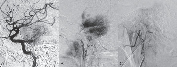
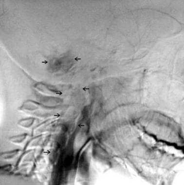
Glomus Jugulare Tumors: Practice Essentials, Relevant Anatomy,  Pathophysiology
Glomus Jugulare Tumors: Practice Essentials, Relevant Anatomy, Pathophysiology

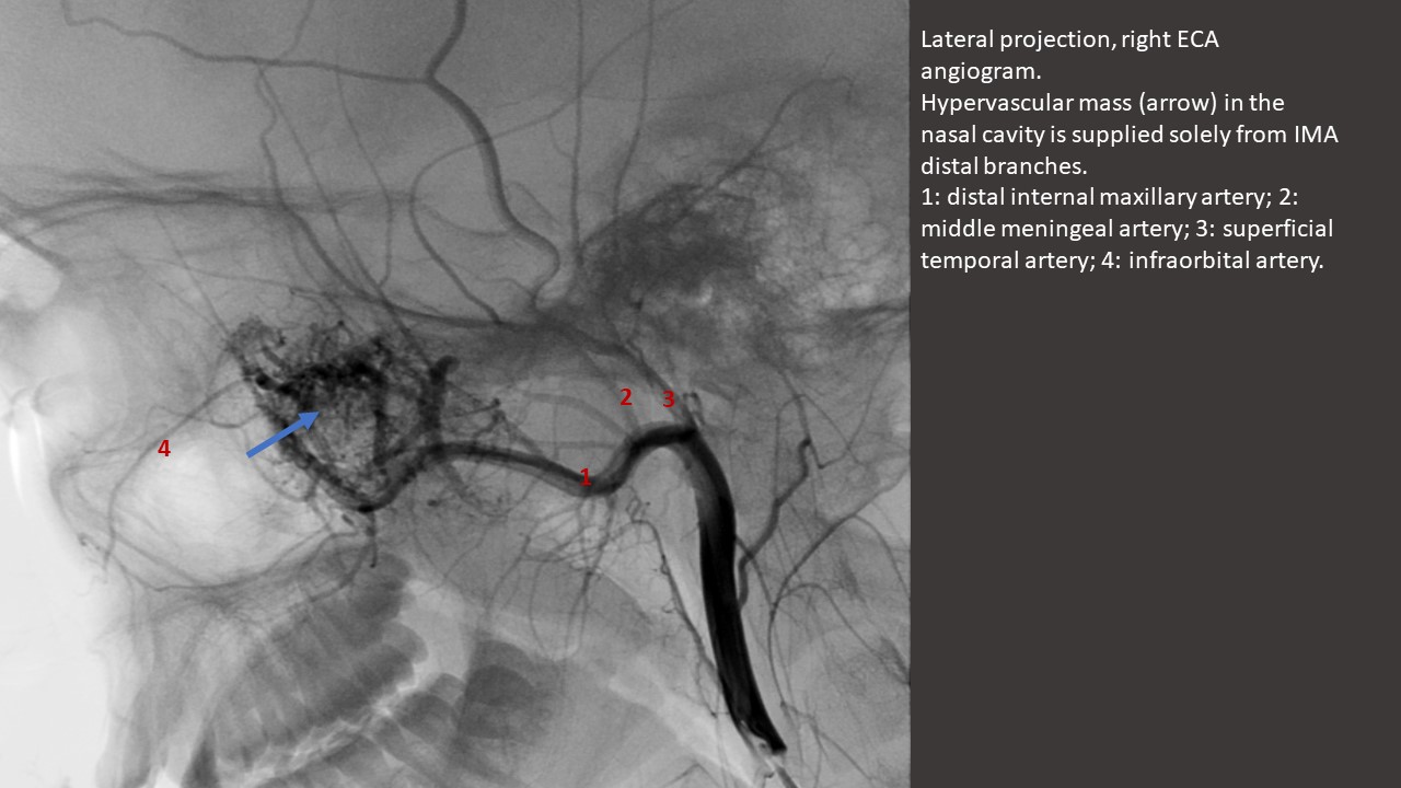
Head, neck, and brain tumor embolization guidelines | Journal of  NeuroInterventional Surgery
Head, neck, and brain tumor embolization guidelines | Journal of NeuroInterventional Surgery

Angiographic appearance of “tumour blush” produced by a large right atrial  myxoma | Heart
Angiographic appearance of “tumour blush” produced by a large right atrial myxoma | Heart

View Image
View Image

Fig 4. | Nasopharyngeal Extension of Glomus Tympanicum: An Unusual Clinical  and Imaging Manifestation | American Journal of Neuroradiology
Fig 4. | Nasopharyngeal Extension of Glomus Tympanicum: An Unusual Clinical and Imaging Manifestation | American Journal of Neuroradiology